Imagining with High-Performance Fluorescence Microscopy Systems
Delivering targeted imaging solutions with precision fluorescence systems tailored for research, diagnostics, and industry.
Advanced Fluorescence Solutions for Research and Industry
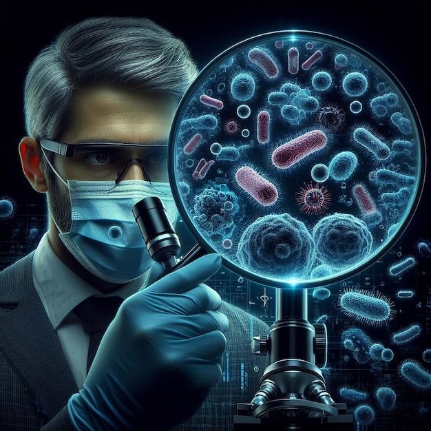
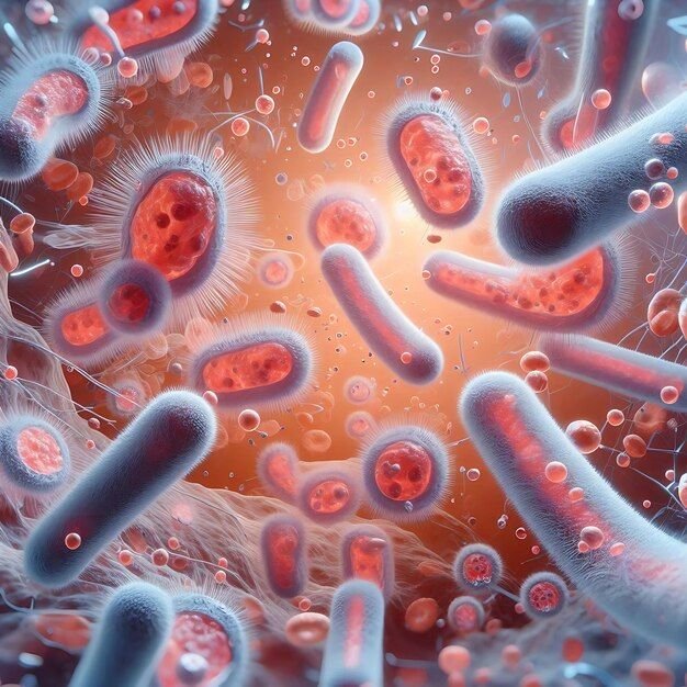
Fluorescence Microscopy enables scientists, engineers, and clinicians to visualize specific structures within complex biological and material samples using targeted fluorescent labeling. By exciting fluorophores with high-intensity light sources, this technique produces high-contrast images that reveal molecular interactions, protein expression, and dynamic cellular processes.
Across North America, businesses and research institutions trust Fluorescence Microscopy to power discoveries and optimize diagnostics. MICROSCOPY AND IMAGING provides integrated, reliable fluorescence solutions tailored to the unique needs of B2B clients in biotechnology, medical research, manufacturing, and environmental monitoring. Our systems combine precision optics with advanced digital platforms to support streamlined workflows and consistent results.With a focus on innovation, product longevity, and client-first support, MICROSCOPY AND IMAGING helps organizations unlock the full potential of fluorescence imaging—from individual lab setups to enterprise-scale deployment.
Core Technologies
In addition to offering products and systems developed by our team and trusted partners for Fluorescence Microscopy, we are proud to carry top-tier technologies from Global Advanced Operations Tek Inc. (GAO Tek Inc.) and Global Advanced Operations RFID Inc. (GAO RFID Inc.). These reliable, high-quality products and systems enhance our ability to deliver comprehensive technologies, integrations, and services you can trust. Where relevant, we have provided direct links to select products and systems from GAO Tek Inc. and GAO RFID Inc.
Hardware
- High-intensity LED or mercury/xenon arc light sourcesare powered reliably using Power Adapters and Converters, ensuring consistent illumination for fluorescent excitation.
- Filter cubes and motorized filter wheels for multiple fluorophoresare enhanced by Motion & Position Sensors to enable precise filter alignment and wavelength control.
- Fluorescence-grade objectives with high numerical aperturesare supported by Surface Roughness Gauges & Testers to ensure ultra-smooth optics essential for high-resolution fluorescence imaging.
- Cooled CMOS or sCMOS cameras for low-noise image captureintegrate well with Optical & Imaging Sensors for enhanced light detection and signal clarity.
- Motorized stages and autofocus systems for precision scanningbenefit from Zigbee End Devices for reliable wireless control in automated microscopy workflows.
- Enclosures for light shielding and live-cell imagingalign with Environmental & Agriculture Sensors to monitor and maintain controlled conditions during live-cell fluorescence observations.
Software
- Real-time image acquisition with channel overlays
- Fluorophore spectral unmixing and quantification tools
- Time-lapse and z-stack capture with 3D reconstruction
- Annotation, measurement, and batch analysis capabilities
- Compatible with AI-based cell recognition and segmentation
Cloud Services
- Centralized image storage and secure backup
- Remote collaboration across research teams
- Integration with image databases and LIMS
- Audit trails for regulatory compliance in diagnostics
Key Features and Functionalities
- Multi-channel fluorescence detection with switchable filters
- Live-cell and fixed-sample imaging
- High-sensitivity detection for weak signals
- Spectral imaging and photobleaching compensation
- Automation-ready with robotic slide loaders and scanners
- Wide compatibility with common dyes like DAPI, FITC, Cy3, and Alexa Fluors.
Integrations
- Digital pathology and telepathology systems
- Real-time diagnostic pipelines
- Automated microscopy workflows and robotic sample handlers
- Image processing platforms like FIJI, Imaris, and MetaMorph
- Hybrid brightfield/fluorescence configurations.
Compatibility
- Compatible with standard microscope frames
- Supports USB 3.0, HDMI, and Ethernet connections
- Interoperable with Windows and macOS-based software
- Supports LIMS and secure cloud-based imaging environments.
Benefits
- Pinpoint accuracy in detecting target structures
- High-throughput screening capabilities
- Real-time visualization and image sharing
- Flexible system configurations for research or industrial QC
- Reliable imaging over long sessions with minimal signal degradation.
Applications
- Gene expression and protein localization studies
- Immunofluorescence and FISH (Fluorescence in Situ Hybridization)
- Cancer diagnostics and biomarker identification
- Pharmaceutical compound screening
- Environmental monitoring of biological agents
- Semiconductor inspection using fluorescent markers.
Industries
- Biomedical Research
- Clinical Diagnostics
- Pharmaceutical R&D
- Environmental Science
- Semiconductor & Nanotech
- Academic and Government Labs.
Relevant Industry Standards
ISO 15189
CLIA Regulations (U.S.)
FDA 21 CFR Part 11
ANSI/AAMI ST79
Health Canada Medical Device Licensing
Case Studies
Cancer Research Center, Texas
A major cancer research institute partnered with MICROSCOPY AND IMAGING to replace outdated fluorescence systems with a fully automated multi-channel setup. The upgrade enabled real-time spectral imaging across 4 fluorescent markers, accelerating their biomarker validation workflow by 40% and increasing image reproducibility across trials.
Diagnostics Manufacturer, California
A diagnostics company specializing in infectious disease testing integrated MICROSCOPY AND IMAGING’s fluorescence platform into their slide-based immunoassay system. Our solution supported rapid image capture and AI-assisted cell counting, enabling scalable production of FDA-compliant test kits with high throughput and consistent accuracy.
Neuroscience Lab, British Columbia
A leading university neuroscience group worked with MICROSCOPY AND IMAGING to develop a custom fluorescence imaging solution for studying calcium ion dynamics in neurons. The implementation of real-time z-stack imaging and automated stage control helped the lab achieve sub-second acquisition across multiple fields, resulting in novel insights into synaptic activity.
Ready to enhance your imaging capabilities with Fluorescence Microscopy?
Contact us today to explore how our systems can support your applications. Our team is available for consultations, technical support, and solution recommendations tailored to your organization.