Precision Imaging with Phase Contrast Microscopy Systems
MICROSCOPY AND IMAGING delivers advanced phase contrast solutions for clear, real-time imaging of live, unstained specimens in research and industry.

Reliable Phase Contrast Solutions for Live, Unstained Sample Analysis
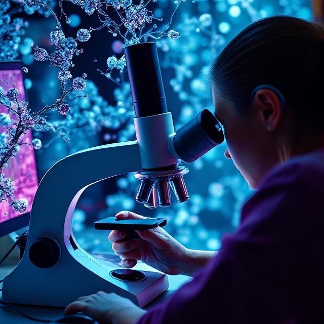
Phase Contrast Microscopy is an essential imaging technique used to visualize transparent, unstained specimens such as live cells, tissues, and microorganisms. By converting subtle differences in light phase into variations in intensity, this method enables users to observe biological dynamics without altering the sample. It offers a non-invasive, cost-effective solution for real-time cellular analysis.
Widely adopted across academic labs, clinical research, and biotech facilities, Phase Contrast Microscopy supports time-lapse imaging and morphological assessment with minimal preparation. MICROSCOPY AND IMAGING supports businesses and laboratories throughout North America with dependable phase contrast solutions tailored to meet specific application needs. As a growing B2B provider, we combine user-friendly systems, expert support, and dependable performance to help organizations elevate their imaging capabilities. Our technology is backed by robust quality assurance, ongoing development, and a commitment to delivering scalable tools for scientific discovery.
Core Components
In addition to offering products and systems developed by our team and trusted partners for Phase Contrast Microscopy, we are proud to carry top-tier technologies from Global Advanced Operations Tek Inc. (GAO Tek Inc.) and Global Advanced Operations RFID Inc. (GAO RFID Inc.). These reliable, high-quality products and systems enhance our ability to deliver comprehensive technologies, integrations, and services you can trust. Where relevant, we have provided direct links to select products and systems from GAO Tek Inc. and GAO RFID Inc.
Hardware
- High-stability compound microscopes with phase contrast optics (enhanced by Optical & Imaging Sensors)
- Phase contrast objectives (typically 10x, 20x, 40x, 100x) with matching phase annuli (supported by Optical & Imaging Sensors)
- Integrated or attachable phase condenser rings (paired with Precision Optical Components under Optical & Imaging Sensors)
- LED or halogen illumination sources with adjustable intensity (compatible with Optical & Imaging Sensors and Device Edgeprocessing under Edge Computing)
- Fine focus and mechanical stage systems (optimized using Motion & Position Sensors)
- Optional digital imaging modules and camera ports (enabled by BLE Gateways, Beacons & Accessories for wireless imaging integration).
Software
- Real-time viewing and image capture
- Frame-by-frame analysis for time-lapse studies
- Image enhancement tools for contrast optimization
- Manual and auto-focus assist tools
- Data annotation and export for presentations or reports
Cloud Services
- Secure storage and archiving of live imaging sessions
- Remote collaboration with real-time screen sharing
- Integration with electronic lab notebooks (ELNs) and data logging systems
- Cloud-based update support and diagnostics
Key Features and Functionalities
- High-contrast visualization of transparent specimens
- Real-time monitoring of cell growth, division, and motility
- Easy switching between brightfield and phase contrast modes
- No staining or sample preparation required
- Suitable for long-term time-lapse imaging
Compact footprint ideal for benchtop or incubator us
Integrations
MICROSCOPY AND IMAGING’s phase contrast systems integrate with:
- Digital slide imaging platforms
- Incubation chambers for live-cell conditions
- Automated stage and focus systems for high-throughput workflows
- Data analysis and quantification software
- Custom lab monitoring tools and cloud-based storage networks
Compatibility
- Compatible with most universal slide formats
- Supports standard mounting frames from Nikon, Olympus, Leica, and Zeiss
- Interfaced with USB and Wi-Fi digital imaging modules
- Operates on Windows and macOS environments
- Compatible with third-party software like ImageJ, Micro-Manager, and Meta morph
Benefits
- Enables observation of live, unstained samples with no prep time
- Preserves biological integrity during extended imaging
- Affordable solution for routine cellular analysis
- Minimal training required for operation
- Flexible for use in academic, clinical, and industrial settings
Applications
- Monitoring of cell cultures and bacterial colonies
- Live tissue analysis in physiological studies
- Quality control in pharmaceutical production
- Microorganism behavior tracking in environmental studies
- Sperm motility testing in fertility labs
- Teaching and student demonstrations in life science education
Industries
- Life Sciences & Biotechnology
- Academic Research & Education
- Clinical Diagnostics & Pathology
- Pharmaceutical Quality Control
- Environmental Testing
- Fertility & Reproductive Health
Relevant Industry Standards
ISO 13485
FDA 21 CFR Part 11
CLIA (Clinical Laboratory Improvement Amendments)
ANSI Z136.1
Health Canada Medical Device Licensing
Case Studies
Cell Therapy Lab, New Jersey
A startup developing regenerative therapies selected MICROSCOPY AND IMAGING’s phase contrast solution for monitoring live stem cell cultures. The non-invasive system allowed the team to track cell viability without introducing stains or sample damage, enhancing QC accuracy while streamlining protocol compliance.
Environmental Testing Center, Oregon
An environmental lab specializing in waterborne pathogen detection partnered with MICROSCOPY AND IMAGING to install modular phase contrast systems. By integrating live-cell observation into their workflow, the lab improved microorganism identification efficiency and reduced sample rejection rates by 25%.
Teaching Hospital, Alberta
A teaching hospital's pathology department adopted MICROSCOPY AND IMAGING's phase contrast microscopes to improve live demonstrations for medical students. Faculty reported improved engagement and understanding of cell structure and dynamics, contributing to more interactive and effective lab sessions.
Want to learn more about how Phase Contrast Microscopy can support your operations?
Contact MICROSCOPY AND IMAGING today for expert guidance, product recommendations, or to schedule a consultation. Let us help you implement a dependable, high-performance imaging solution tailored to your needs.